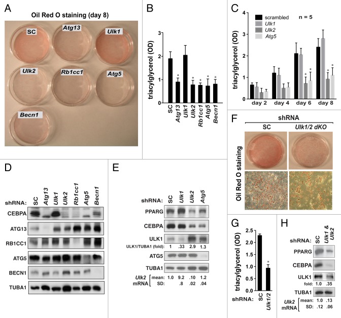
Figure 1.Ulk1 is not required for adipogenesis in 3T3-L1 cells. (A) Knockdown of autophagy genes, except Ulk1, suppresses adipogenesis. 3T3-L1 cells were transduced by shRNAs specific to each autophagy gene. As a control, 3T3-L1 cells were transduced by scrambled (SC) shRNA. The shRNA-transduced cells were induced to be differentiated into adipocytes in medium containing methylisobutylxanthine, dexamethasone, and insulin as described previously.37 At day 8, cells were stained with Oil Red O. (B) Quantitative analysis of intracellular triglyceride content at day 8. Triglycerides were extracted from Oil Red O-stained cells with isopropanol, and the contents were analyzed by measuring the optical density at 490 nm. Values were normalized by protein concentration and presented as mean ± SD *P < 0.01 relative to shRNA-SC cells. (C) Quantitative analysis of intracellular triglyceride content over the period of differentiation. Mean ± SD from 3 independent experiments. *P < 0.01 relative to scrambled control cells. (D) western blot analysis of CEBPA and the gene knockdown in shRNA-transduced cells. (E) ULK1 and ULK2 reciprocally regulate their expression in adipocytes. The expression levels of ULK1, ULK2, and ATG5 in shRNA-transduced adipocytes at day 8 were analyzed by western blotting and quantitative real time RT-PCR. (F) Knockdown of both Ulk1 and Ulk2 suppresses adipogenesis to a similar extent as knockdown of Ulk2 alone. (G) Quantitative analysis of intracellular triglyceride content over the period of differentiation. Values are mean ± SD from 5 independent experiments. *P < 0.01 relative to control cells. (H) western blot analysis of the effects of knocking down both Ulk1 and Ulk2 on the expression of PPARG and CEBPA.

An official website of the United States government
Here's how you know
Official websites use .gov
A
.gov website belongs to an official
government organization in the United States.
Secure .gov websites use HTTPS
A lock (
) or https:// means you've safely
connected to the .gov website. Share sensitive
information only on official, secure websites.
