
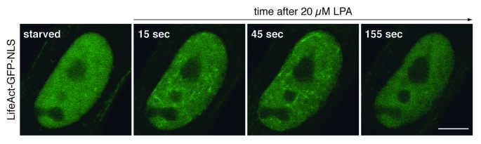
Figure 1. Signal-responsive nuclear actin dynamics. Live NIH3T3 cells expressing the actin probe LifeAct-GFP-NLS were monitored before and during stimulation with 20 µM lysophosphatidic acid (LPA). Prior to analysis, cells were transiently transfected with a plasmid encoding LifeAct-GFP-NLS and kept in serum-free medium for 24 h. Confocal microscopic images (1 frame every 10 s) reveal the distribution of LifeAct-GFP-NLS at indicated time points. Note that LPA-stimulation triggers an immediate and transient formation of nuclear actin filaments, which become visible by the decoration with LifeAct-GFP-NLS. Scale bar, 10 µm.