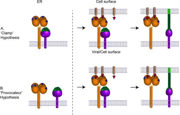
Figure 2. Schematic diagram illustrating the ‘Clamp’ hypothesis and the ‘Provocateur’ hypothesis.
(A) In the ’Clamp’ hypothesis the attachment protein HN, (H or G) and the F protein interact during their transport through the exocytic pathway and at the cell or viral surface dissociate upon receptor engagement with the attachment protein, allowing the F protein to trigger in a manner analogous to a loaded spring. (B) In the ‘Provocateur’ hypothesis, the attachment protein and the F proteins do not associate inside the cell but do associate at the cell or viral surface, and dissociate after the attachment protein engages with the receptor. The protein-protein interaction between HN (H, G) and F triggers the F protein. The receptor binding protein is illustrated in orange as a globular head linked, via a flexible linker, to a four helix bundle stalk. The receptor binding site is shown as a blue triangle. The F protein is illustrated as a purple trimer with the domain that refolds in green. The receptor molecule is illustrated as a light brown cylinder with a red triangle as the attachment point. Potential changes in the HN/H/G stalk on receptor engagement are not illustrated.

