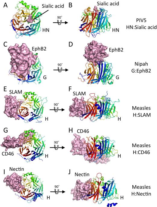
Figure 3. Receptor complexes of paramyxovirus attachment glycoprotein receptor binding domains.
For all panels, the receptor binding domains (RBDs) are shown in cartoon format, colored in a rainbow from N (blue) to C (red). The receptors are shown in solid surface format colored light pink. The RBDs all show a characteristic 6-bladed beta-propeller domain that is observed in sialidases and neuraminidases, although the Nipah virus G and measles virus H proteins do not bind sialic acid as their receptors. Pairs of panels are shown with two views of each receptor complex, related by a 90° rotation as indicated. (A, B) Two views of the PIV5 HN:sialylalactose complex (PDB ID code 1Z4X). (C, D) Two views of the Nipah virus G:EphB2 complex (PDB ID code 2VSM). (E, F) Two views of the measles virus H:SLAM complex (PDB ID code 3ALZ). (G, H) Two views of the measles virus H:CD46 complex (PDB ID code 3INB). (I, J) Two views of the measles virus H:nectin complex (PDB ID code 4GJT).

