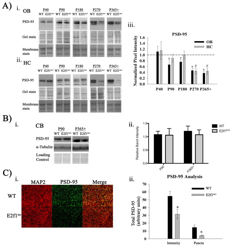
Figure 5.
Age-dependent reduction in PSD-95 expression in hippocampus and olfactory bulbs. A) Immunoblots of PSD-95 expression in OB (i.) and HC (ii.) across age groups. Coomassie-stained gels and fast green-stained membranes are shown as loading controls. (iii.) Quantification of the densitometry analysis displayed as a ratio of the E2f1tm1 to the WT. (N=6. *, OB; #, HC) B) (i.) Immunoblots of PSD-95 expression in cerebellum across two representative age groups P90 and P365+. (ii.) Quantification of the densitometry analysis (N=5 P90, N=6 P365+). C) (i.) Representative images of coronal WT and E2f1tm1 P365+ hippocampal sections immunolabeled with MAP2 (red) and PSD-95 (green) captured at 400x. (ii.) Quantification of the total PSD-95 pixel intensity and the intensity-saturated PSD-95 puncta in the WT and E2f1tm1 (N=3 for each genotype, 8 sections each, * Student’s t-test; α ≤ 0.05. All data are represented as mean ± SEM.

