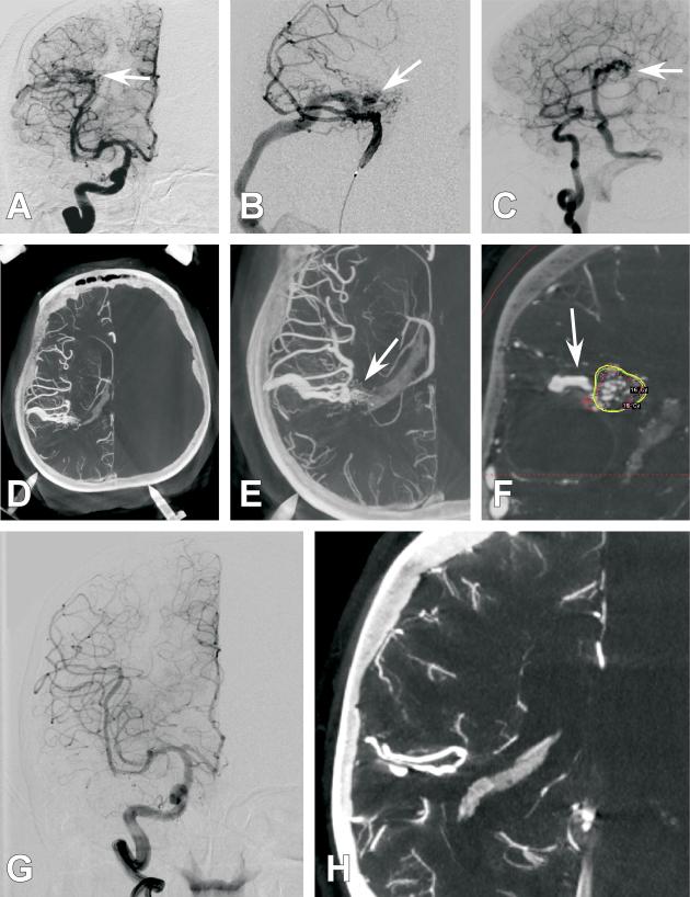
Figure 1. Case 4.
A 61-year old female presented with a right parietal hemorrhage, generalized tonic clonic seizure, and left hemiparesis. She was found to harbor an AVM at the site of hemorrhage and was treated with GKR 1 month after presentation. Several diagnostic cerebral angiogram images are presented. A.) Anterio-posterior (AP) projection of an internal carotid artery (ICA) injection. Poor detailed arterial, nidal, and venous resolution of the AVM are noted (White arrow). B.) Anterio-posterior (AP) projection of a selective middle cerebral artery (MCA) injection. Poor detailed arterial, nidal, and venous resolution of the AVM are noted (White arrow). C.) 3DRA right ICA injection with a right parietal AVM (white arrow) but with continued limited arterial, nidal, and venous anatomy. D.) Axial CBCT-A reconstruction with patient in the Leksell head frame. The anterior posts and posterior pins of the frame are seen. A right parietal AVM is visualized. E.) Magnified axial CBCT-A reconstruction. Excellent resolution of the complex arterial and venous structure of the AVM are noted (White arrow). F.) Screen shot image of a coronal CBCT-A reconstruction uploaded for planning on the Leksell stereotactic planning computer. The radiated field is demonstrated by the encircled areas at different radiation isodoses. Note that the draining vein is easily resolved and left out of the radiation field (White arrow). G.) 2-year follow-up AP projection of an ICA injection demonstrating complete obliteration of the previously seen AVM. H.) 2-year follow-up magnified axial CBCT-A reconstruction demonstrating complete obliteration of the previously seen AVM.

