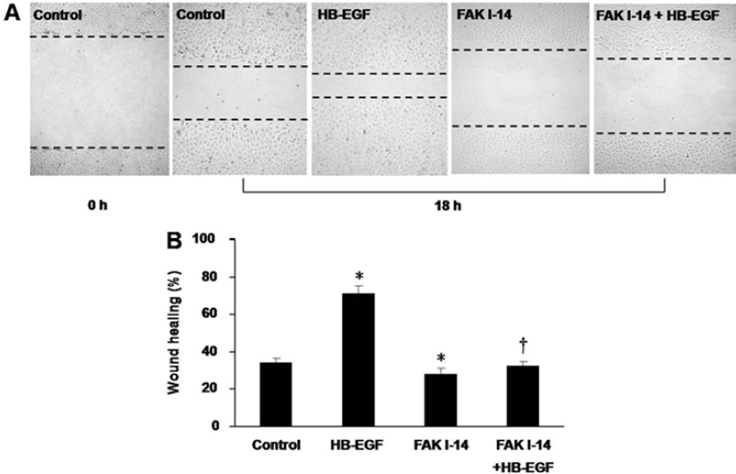
Figure 2.
RIE-1 cell migration after scrape wounding in the presence of p-FAK inhibition. (A) Representative images of RIE-1 cells 18 h after scrape wounding. From left to right: 0 h after scrape wounding of untreated RIE-1 cells; 18 h after wounding of untreated cells, HB-EGF-treated cells, FAK1-14-treated cells and cells treated with FAK1-14 plus HB-EGF. The black dotted lines present the wound edges at 18 h. Scale bar = 200μm. (B) Effect of p-FAK inhibition on RIE-1 cell migration 18 h after scrape wounding. *p<0.05, vs. control; † p<0.05 vs. HB-EGF.

