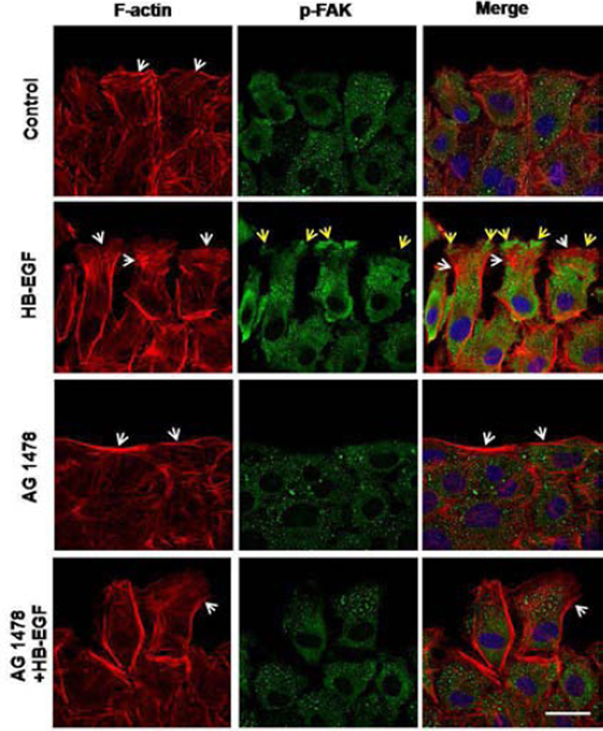
Figure 3.
Distribution of F-actin filaments and p-FAK after scrape wounding. Shown are representative immunofluorescent images of F-actin filaments (red) and p-FAK (green) in migrating RIE-1 cells 18 h after scrape wounding. At the wound edges, the white arrows indicate lamellipodia, and the yellow arrows indicate polarization of p-FAK to the foremost edge of the migrating RIE-1 cells. Scale bar = 20 μm.

