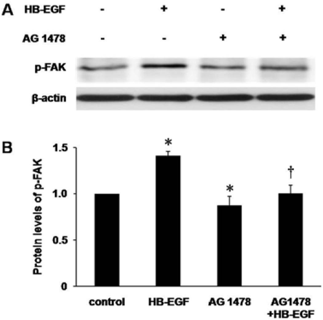
Figure 4.
p-FAK protein levels in RIE-1 cells as determined by Western blotting. (A) Western blotting of p-FAK 18 h after scrape wounding. (B) Densitometric quantification of protein levels of p-FAK. The protein level of p-FAK in control group was defined as 1.0. *p<0.05 vs. control; † p<0.05 vs. HB-EGF.

