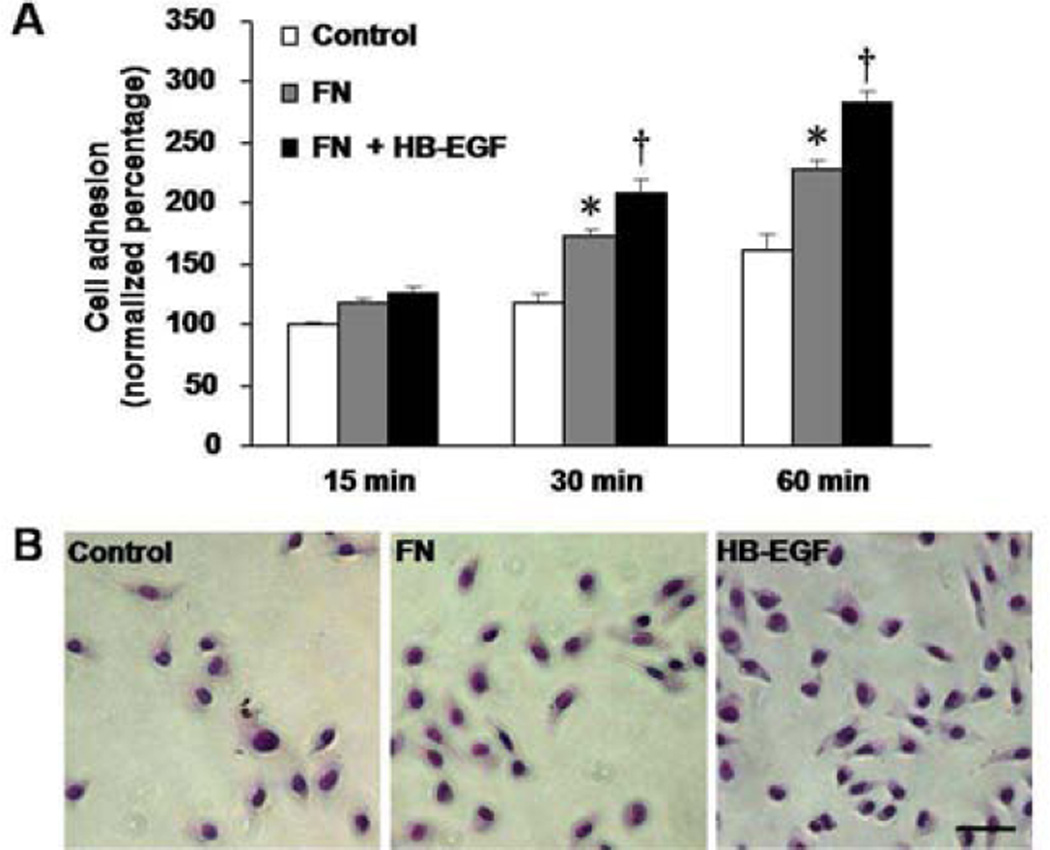
Figure 5.
RIE-1 cell adhesion on fibronectin (FN). (A) RIE-1 cells adhesion on FN is shown at different time points. *p<0.05vs. control cells plated in the absence of FN; † p<0.05 vs. cells grown on FN in the absence of HB-EGF. (B) Representative images ofRIE-1 cell adhesion after 60 min of incubation. Scale bar = 30 μm.

