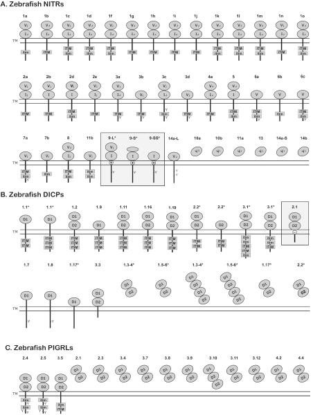
Figure 2. Predicted structures of zebrafish NITR, DICP, and PIGRL proteins.
Sequences have been reported (Haire et al. 2012;Kortum et al. 2014;Yoder et al. 2004;Yoder et al. 2008). Proteins are organized by inhibitory, activating, functionally ambiguous and secreted forms. Activating receptors are boxed. The Ig domains of (A) NITR (V and I), (B) DICP (D1 and D2) and (C) PIGRL (D1 and D2) proteins are indicated. Ig domains that include a joining (J) or J-like (j) sequence are labeled with a subscript J or j. Cytoplasmic ITIMs, ITIM-like (itim) sequences and tyrosines as well as charged residues (⊕ or ⊖) within transmembrane domains are labeled. Proteins encoded by splice variants are indicated with asterisks (*). This is not meant to be a complete catalog of proteins from these families as additional proteins sequences and structures will likely be identified in the future.

