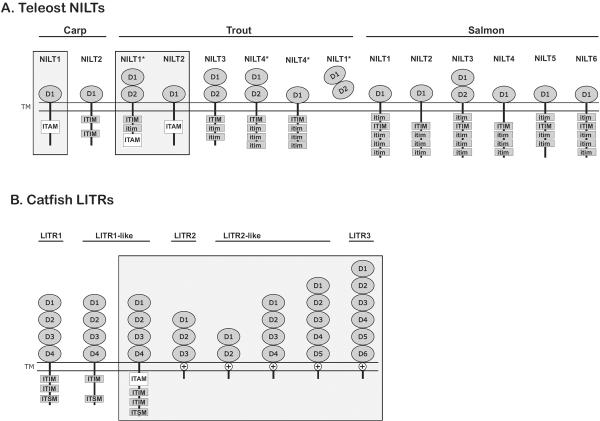
Figure 3. Predicted structures of teleost NILT and LITR proteins.
Sequences have been reported (Montgomery et al. 2011;Ostergaard et al. 2009;Ostergaard et al. 2010;Stet et al. 2005). Proteins are organized numerically: activating receptors are boxed. The Ig domains of (A) NILT (D1 and D2) and (B) LITR (D1, D2, D3, D4, D5 and D6) proteins are indicated. Cytoplasmic ITAMs, ITIMs, ITIM-like (itim) sequences and tyrosines as well as charged residues (⊕) within transmembrane domains are labeled. Proteins encoded by splice variants are indicated with asterisks (*). This is not meant to be a complete catalog of proteins from these families as additional proteins sequences and structures will likely be identified in the future.

