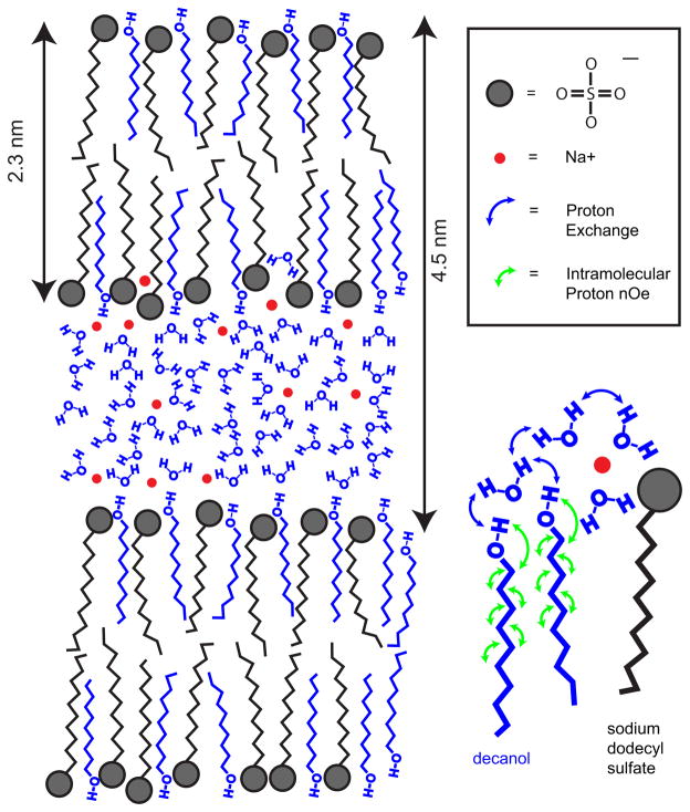
Figure 1.
Conceptual diagram of the LLC phase formed by hydrophobic bilayers of SDS (black) and decanol (blue) chains that encapsulate polar water layer (blue) and sodium ions (red). The distances are given for water content of CW = 45% as measured by small angle X-ray scattering (SAXS) experiments Ref. (34). Blue color indicates LLC components (decanol and water) participating in MT. The insert on the right focuses on a sub-set of the polar / non-polar interface to reflect the MT mechanism between decanol and water protons consistent with our experimental findings, i.e., proceeding via hydroxyl proton exchange (blue arrows) and aliphatic proton nOe (green arrows). Terminal chain segments of decanol are excluded from MT due to probable large amplitude motions.

