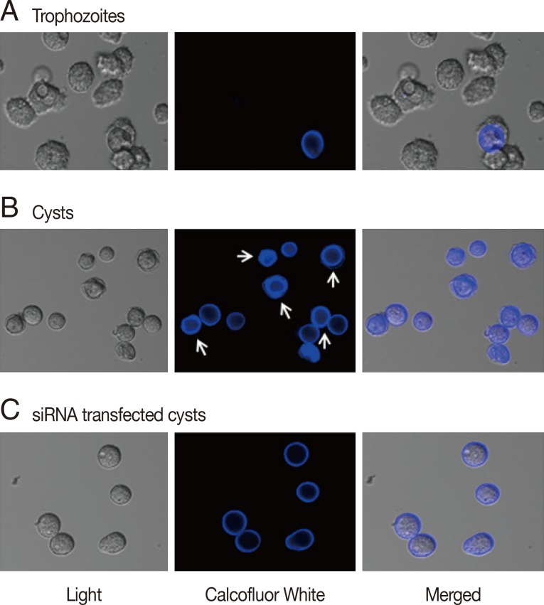
Fig. 2.
Cyst wall formation as detected by calcofluor white staining. (A) Cellulose was not present in trophozoites by calcofluor white staining. (B) At day 3 after the induction of encystation, young cysts and mature cysts (arrows) were observed. (C) The majority of cellulose synthase siRNA transfected cells were young or immature cysts.

