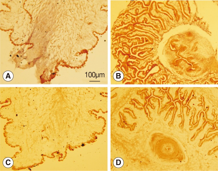
Fig. 2.
Analysis of wnt4 expression by in situ hybridization. (A, B) Antisense probes were used. (C, D) Sense probes were used. (A, C) Scolex evaginated cysticercus. (B, D) Scolex invaginated cysticercus. The expression of wnt4 was deeply stained from the posterior end along the primary body axis of cysticercus as shown in A.

