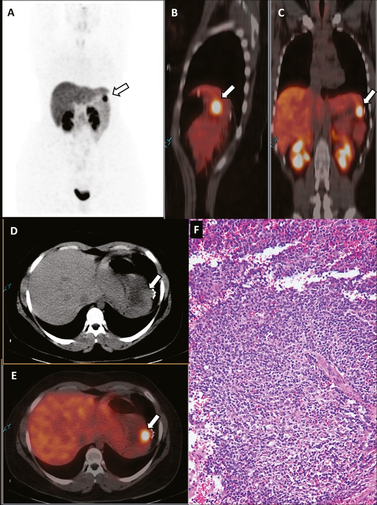
A 48-year-old female patient underwent splenopancreasectomy for a 4-cm pancreatic neuroendocrine tumor (pNET), grade G2, located in the pancreatic tail. One year after surgery, the patient presented an increased serum level of the tumor marker chromogranin A (value: 160 U/l). Therefore, she underwent somatostatin receptor PET/CT using gallium-68-DOTATOC for restaging. This imaging method showed a focal area of increased radiopharmaceutical uptake corresponding to a 2.5-cm nodule located in the left superior abdomen near a clip from the previous surgery, suggesting a possible relapse of pNET (Fig. 1). Based on this PET/CT finding, the patient underwent ultrasonography-guided core biopsy of this nodule. Histology did not reveal findings suggestive of pNET but identified spleen tissue most likely caused by splenosis accidentally seeded at the previous operation. It is likely that the increased serum level of the tumor marker chromogranin A was due to the chronic proton-pump inhibitors use.
Fig. 1.
A 48-year-old female patient previously treated with splenopancreasectomy for a 4-cm pNET, grade G2, located in the pancreatic tail underwent somatostatin receptor PET/CT for restaging because of an increase in the chromogranin A serum levels (value: 160 U/l). Gallium-68-DOTATOC was injected (activity: 140 MBq). Images were acquired 1 h after radiopharmaceutical injection. Maximum standardized uptake values (SUVmax) were used to measure the radiopharmaceutical uptake semi-quantitatively. Somatostatin receptor PET (a), sagittal (b) and coronal (c) PET/CT, axial CT (d) and PET/CT (e) images showed a focal area of increased radiopharmaceutical uptake (SUVmax: 13) corresponding to a 2.5-cm nodule located in the left superior abdomen (arrows) near a clip from the previous surgery, suggesting a possible relapse of pNET. Based on this PET/CT finding, the patient underwent ultrasonography-guided core biopsy of this nodule. Histology did not reveal findings suggestive of NET but identified spleen tissue (f), most likely caused by splenosis accidentally seeded at the previous operation
Somatostatin receptor PET/CT is an accurate imaging method for staging and restaging pNET, presenting high sensitivity and specificity in this setting [1–7]. Nevertheless, possible sources of false-negative and -positive findings with this method should be taken into account [1]. Inflammatory lesions represent the most frequent causes of false-positive findings for pNET at somatostatin receptor imaging because inflammatory cells may overexpress somatostatin receptors on their cell surface [8, 9].
In our case, we showed that splenosis may represent a possible cause of false-positive findings for pNET relapse due to the physiological uptake of somatostatin analogs by the spleen tissue [10, 11].
Acknowledgments
Conflict of Interest
Giorgio Treglia, Luca Giovanella, Barbara Muoio and Carmelo Caldarella declare that they have no conflicts of interest.
Funding
None.
References
- 1.Treglia G, Castaldi P, Rindi G, et al. Diagnostic performance of gallium-68 somatostatin receptor PET and PET/CT in patients with thoracic and gastroenteropancreatic neuroendocrine tumours: a meta-analysis. Endocrine. 2012;42:80–87. doi: 10.1007/s12020-012-9631-1. [DOI] [PubMed] [Google Scholar]
- 2.Treglia G, Cason E, Fagioli G. Recent applications of nuclear medicine in diagnostics (first part) Ital J Med. 2010;4:84–91. doi: 10.1016/j.itjm.2010.03.001. [DOI] [Google Scholar]
- 3.Rufini V, Baum RP, Castaldi P, et al. Role of PET/CT in the functional imaging of endocrine pancreatic tumors. Abdom Imaging. 2012;37:1004–1020. doi: 10.1007/s00261-012-9871-9. [DOI] [PubMed] [Google Scholar]
- 4.Oh J-R, Kulkarni H, Carreras C, et al. Ga-68 Somatostatin receptor PET/CT in von Hippel-Lindau disease. Nucl Med Mol Imaging. 2012;46:129–133. doi: 10.1007/s13139-012-0133-0. [DOI] [PMC free article] [PubMed] [Google Scholar]
- 5.Treglia G, Salomone E, Petrone G, et al. A rare case of ectopic adrenocorticotropic hormone syndrome caused by a metastatic neuroendocrine tumor of the pancreas detected by 68Ga-DOTANOC and 18F-FDG PET/CT. Clin Nucl Med. 2013;38:e306–e308. doi: 10.1097/RLU.0b013e318279ec68. [DOI] [PubMed] [Google Scholar]
- 6.Treglia G, Inzani F, Campanini N, et al. A case of insulinoma detected by 68Ga-DOTANOC PET/CT and missed by 18F-dihydroxyphenylalanine PET/CT. Clin Nucl Med. 2013;38:e267–e270. doi: 10.1097/RLU.0b013e31825b222f. [DOI] [PubMed] [Google Scholar]
- 7.Treglia G, Plastino F, Campitiello M. Staging and treatment response evaluation in a metastatic neuroendocrine tumor of the pancreas with G2 grading: insights from multimodality diagnostic approach by F-18-FDG and Ga-68-DOTANOC PET/CT. Endocrine. 2013;43:729–731. doi: 10.1007/s12020-012-9858-x. [DOI] [PubMed] [Google Scholar]
- 8.Castaldi P, Rufini V, Treglia G, et al. Impact of 111In-DTPA-octreotide SPECT/CT fusion images in the management of neuroendocrine tumours. Radiol Med. 2008;113:1056–1067. doi: 10.1007/s11547-008-0319-9. [DOI] [PubMed] [Google Scholar]
- 9.Treglia G, Farchione A, Stefanelli A, et al. Masking effect of chronic pancreatitis in the interpretation of somatostatin receptor positron emission tomography in pancreatic neuroendocrine tumors. Pancreas. 2013;42:726–728. doi: 10.1097/MPA.0b013e3182750ea0. [DOI] [PubMed] [Google Scholar]
- 10.Shetty D, Lee Y-S, Jeong JM. 68Ga-labeled radiopharmaceuticals for positron emission tomography. Nucl Med Mol Imaging. 2010;44:233–240. doi: 10.1007/s13139-010-0056-6. [DOI] [PMC free article] [PubMed] [Google Scholar]
- 11.Kulkarni HR, Prasad V, Kaemmerer D, et al. High uptake of (68)Ga-DOTATOC in spleen as compared to splenosis: measurement by PET/CT. Recent Results Cancer Res. 2013;194:373–378. doi: 10.1007/978-3-642-27994-2_19. [DOI] [PubMed] [Google Scholar]


