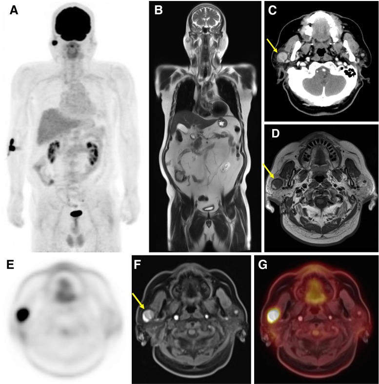
Fig. 1.
Warthin tumor of right parotid gland in a 60-year-old man. a MIP image of whole-body 18F-FDG PET. b Coronal HASTE T2-weighted image of the whole body. c Axial image of contrast-enhanced CT of the head. d Axial image of Gd-contrast enhanced T1-weighted image of the head. e Axial dedicated PET image of the head. f Axial VIBE image of the head. e Axial integrated PET image combined with axial VIBE image

