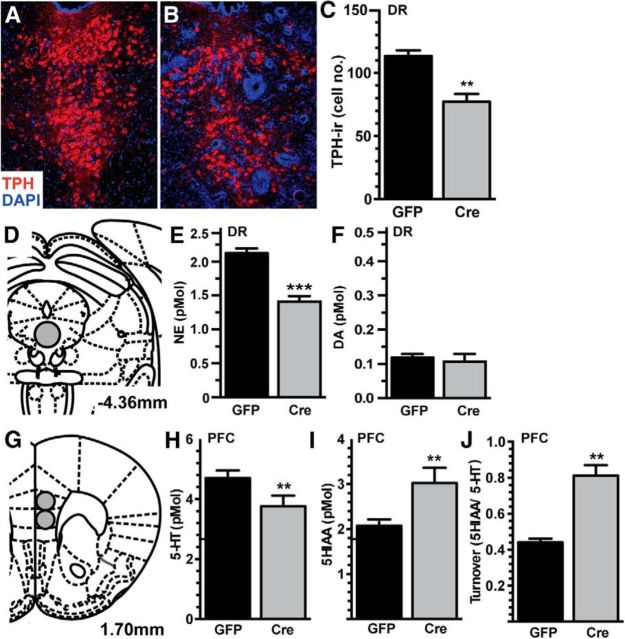
Figure 6.
IL-1β-mediated neuroinflammation in the DR results in serotonergic hypofunction. A, B, Immunofluorescence of TPH-immunoreactivity was used to identify serotonergic neurons (red), counterstained with the nuclear stain DAPI (blue), of AAV-GFP-infused (A) and AAV-Cre-infused (B) DR 8 weeks following AAV infusion (n = 4). C, Quantification revealed fewer TPH-immunoreactive cells in AAV-Cre-infused animals. D–F, HPLC analysis of biogenic amines of the DR (D) revealed less NE (E) but no change in DA (F). G–J, In the PFC (G), AAV-Cre-infused animals had less 5-HT (H), increased levels of the metabolite (I, 5HIAA), and an overall increase in neurotransmitter turnover (J, 5HIAA/5-HT ratio). Data are presented as mean ± SEM, n = 6–7; **p < 0.01, ***p < 0.0001 following t test comparing AAV-GFP and AAV-Cre infusion.

