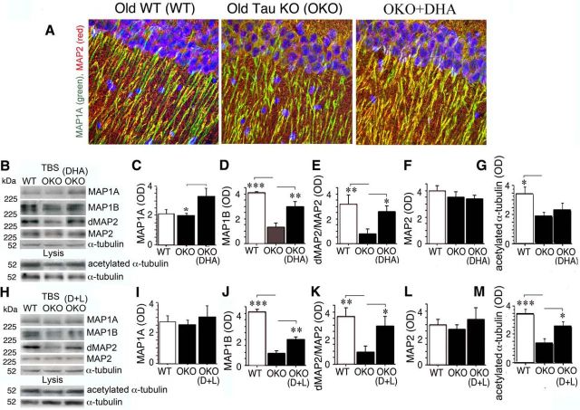
Figure 5.
DHA-supplemented diets partially rescued MAP1A, MAP1B, and MAP2 deficits and microtubule stability in OKO. A, Confocal microscopy demonstrated that DHA diet increased high staining intensity of MAP1A (green) in the dendritic field of the hippocampal CA1 (stratum radiatum) of OKO relative to OKO on control diet so that it better resembled the pattern seen in WT mice. B–M, WB analysis showed that DHA (p < 0.05; B), but not D+L (p > 0.05; I) diet significantly increased MAP1A in OKO compared with mice on control diet. Both DHA and D+L partially restored the loss of total MAP1B in OKO (p < 0.01; D, J). Dephospho MAP2, specific for active MAP2, was also significantly elevated by both treatments (p < 0.05; E, K), whereas total MAP2 did not change (p > 0.05; F, L). Acetylated α-tubulin was significantly reduced in OKO (p < 0.05, G; p < 0.001, M) from two independent animal experiments with different groups raised at different times, whereas the reduction of acetylated α-tubulin was significantly improved by D+L (p < 0.05; H, M), but not by DHA alone (p > 0.05; B, G).

