Figure 1.
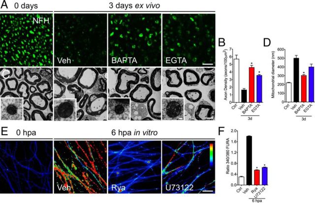
Differential effects of intracellular and extracellular calcium chelators on axonal mitochondria. Sciatic nerve explants from wild-type mice were incubated in vehicle solution (Veh), BAPTA-AM (100 μm), or EGTA (6 μm) for 3 d and analyzed by EM and immunofluorescence. A, Transverse sections of nerve explants stained for NFH. The NFH signal decreases considerably after 3 d in vehicle solution. In nerves treated with BAPTA or EGTA, strong preservation of NFH is observed. Scale bar, 20 μm. B, Quantification of axons positive for NFH in explant cross-sections as shown in A, expressed as axons per 100 μm2. A statistically significant difference in axonal density is seen after either of the calcium chelators treatment for 3 d (n = 3 per group; *p < 0.05 by Student's t test compared with 3 d of vehicle; error bars indicate SEM). C, At the ultrastructural levels, non-injured nerves show conserved axons and compacted myelin sheaths. Conversely, nerves incubated for 3 d in vehicle solution have degenerated axons and collapsed myelin sheaths. Only chelation of intracellular calcium using BAPTA-AM prevents mitochondrial swelling (inset). D, Quantification of mitochondrial diameters from transverse sections analyzed by EM in vehicle-, BAPTA-AM-, and EGTA-treated nerve explants (n = 3 per group; *p < 0.05 by Student's t test compared with 3 d of vehicle; error bars indicate SEM). Scale bars: C, 20 μm; inset, 300 nm. E, Embryonic DRG explants in vitro were axotomized and treated with Rya (5 μm) or U73122 (2 μm). At 6 hpa, cultures were treated for 30 min with fura-2. Treatment with either Rya or U73122 protects axons from intra-axonal calcium increase. The arbitrary color scale at the right indicates relative levels of calcium, with red and blue representing the high and low ends of calcium levels, respectively. Scale bar, 50 μm. F, Quantification of calcium increase by fluorescence intensity of ratio 340/380 (for details, see Materials and Methods). Either Rya (5 μm) or U73122 (2 μm) decreases calcium signal at 6 hpa (n = 9, 3 images per sample, 3 samples per condition, 3 independent experiments; *p < 0.05 by Student's t test compared with control; error bars indicate SEM).

