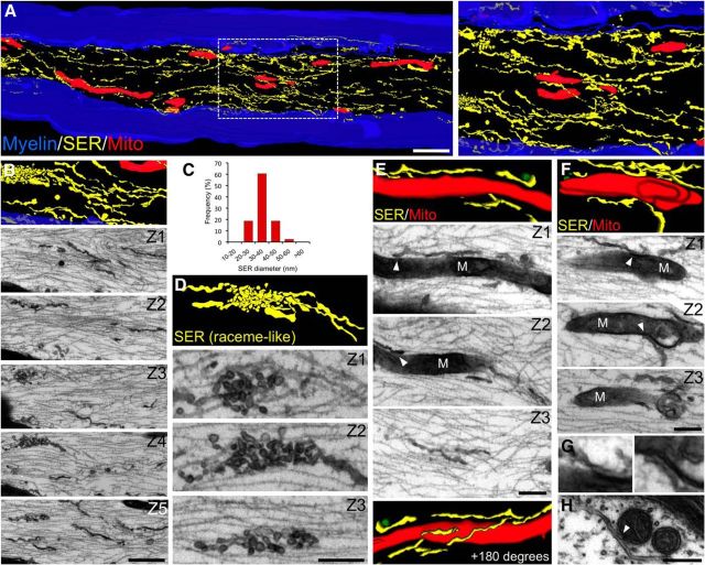
Figure 4.
3D axon reconstruction shows close appositions between mitochondria and ER organelles in peripheral axons. Sciatic nerves from wild-type mice were fixed, and serial sections at ∼80 nm intervals were obtained (see Materials and Methods). After section alignment, axonal membrane structures and mitochondria were reconstructed from myelinated fibers and rendered. Reticular structures are colored yellow, mitochondria in red, and the myelin sheath in blue. Selected micrographs from contiguous sections are included for each rendering. A, 3D reconstruction shows an extensive smooth endoplasmic reticulum (SER) network and elongated mitochondria of varying sizes. The right shows a higher magnification of the boxed area from the left. C, Quantification of axonal SER diameter. Different types of morphological configurations of the SER network are observed, including fibrillar (B) and specializations in the form of raceme-like structures (D). E, F, Regions of close proximity between mitochondria (M) and SER in the axonal compartment (see arrowheads in individual sections). G, Higher magnification of regions indicated by arrowheads in F. H, Cross-section of a myelinated axon in which a SER structure is closely apposed to two mitochondria. Scale bars, A, 1 μm; B, 500 nm; D, 250 nm; E, F, 200 nm; H, 200 nm.

