Figure 5.
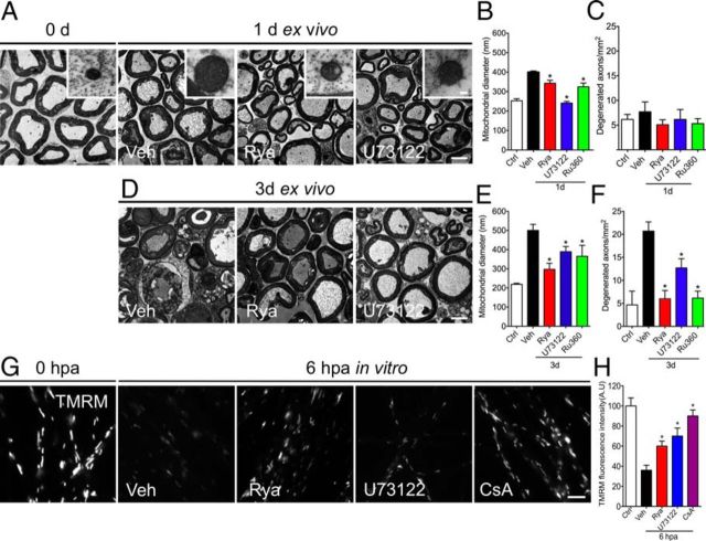
Pharmacological inhibition of ER calcium channels protects against mitochondrial swelling. A, D, Representative electron micrographs of non-damaged neurites (0 h) and explants incubated for 1 or 3 d in vehicle (Veh) or ER calcium channels blockers Rya and U73122 or Ru360. Inhibition of ER channels or the mitochondrial uniporter prevents mitochondrial swelling at 24 hpa (A) and 72 hpa (D). Scale bars, 20 μm; inset, 300 nm. B, E, Quantification of mitochondrial diameters from transverse sections analyzed by EM in vehicle-, Rya-, U73122-, and Ru360-treated nerve explants. Mitochondrial swelling was inhibited by Rya, U73122, and Ru360 at 1 and 3 d (n ≥ 45 mitochondria/nerve over 3 separate experiments; *p < 0.05 by Student's t test compared with vehicle). C, F, Quantification of degenerated axons from transverse sections analyzed by EM in vehicle-, Rya-, U73122-, and Ru360-treated nerve explants. At 3 d, Rya, U73122, and Ru360 inhibit axonal degeneration of nerve explants (n = 3 per group; *p < 0.05 by Student's t test compared with 1 d (C) and 3 d (F) of vehicle; error bars indicate SEM). G, H, Embryonic DRGs were axotomized and treated with vehicle, Rya (5 μm), or U73122 (2 μm). After 6 h, they were stained with TMRM, a mitochondrial membrane potential vital dye. G, TMRM fluorescence in non-injured DRG neurites or 6 hpa DRGs treated with vehicle solution, Rya, U73122, or CsA. Rya, U73122, and CsA inhibit the loss of TMRM fluorescence after axotomy. Scale bar, 50 μm. H, Quantification of TMRM fluorescence in each condition normalized to control (n = 9, 3 images per sample, 3 samples per condition, 3 repetitions; *p < 0.05 by Student's t test compared with vehicle; error bars indicate SEM).

