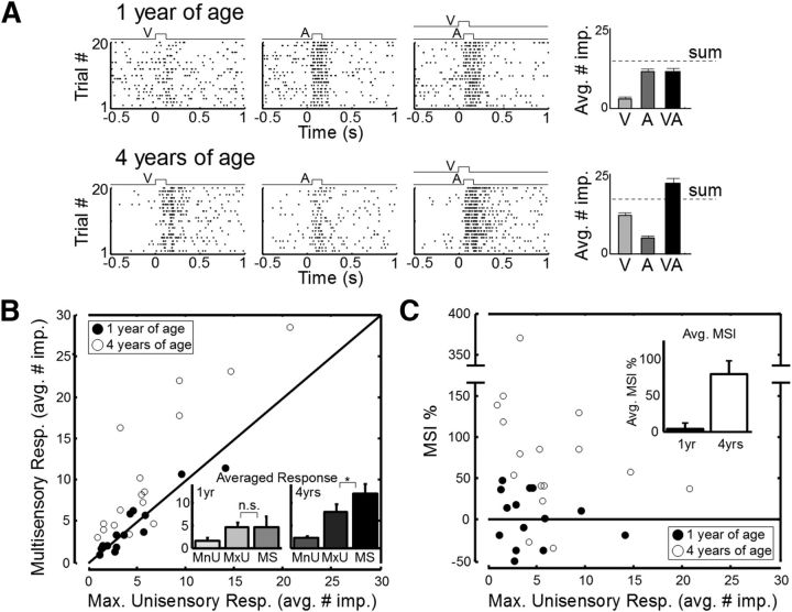
Figure 3.
Changes in multisensory enhancement capabilities were also observed in SC physiology. A, Recordings from exemplar multisensory SC neurons ipsilateral to early cortical deactivation at 1 year of age (top) and 4 years of age (bottom). Responses to a cross-modal (visual–auditory) stimulus were no more robust than to the most effective component stimulus at 1 year but were significantly enhanced at 4 years. Bar graphs on the right represent the magnitude of each response (average no. of stimulus-elicited impulses). Error bars indicate SEM. B, The patterns of enhancement observed in the individual neurons in A were reflected in populations of ipsilateral SC multisensory neurons recorded at 1 year of age (filled circles, n = 14) and 4 years of age (open circles, n = 16) in animals given these treatments. Plotted is the multisensory response magnitude (average no. of impulses) versus the maximum of the component unisensory response magnitudes for each neuron. Inset, Averaged response magnitudes across each population for three categories: the minimum unisensory component response (MnU), the maximum unisensory response (MxU), and the multisensory response (MS). C, The relationship between MSI and the maximum unisensory response for the neurons recorded in the two age groups. Error bars indicate SEM. Inset, Average MSI across these two cohorts. As in the behavioral tests, multisensory integration capabilities that were compromised at 1 year of age had developed at 4 years of age.

