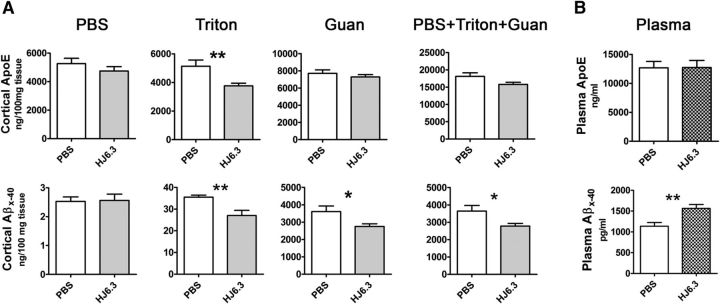
Figure 4.
Effects of the anti-apoE antibody HJ6.3 on Aβ and apoE levels in the brain and blood. A, The cortices of the HJ6.3- or PBS-treated mice were homogenized in PBS, followed by 1% Triton X-100 and 5 m guanidine (Guan). ApoE and Aβx-40 levels in the brain tissue lysates in the PBS, Triton X-100, and Guan fractions were measured by ELISA. The level of apoE and Aβx-40 in all fractions combined is also shown (n = 10–18/group). B, Plasma apoE and Aβx-40 levels in HJ6.3- and PBS-treated animals were measured by ELISA (n = 15–18/group; *p < 0.05; **p < 0.01).

