
Figure 2.
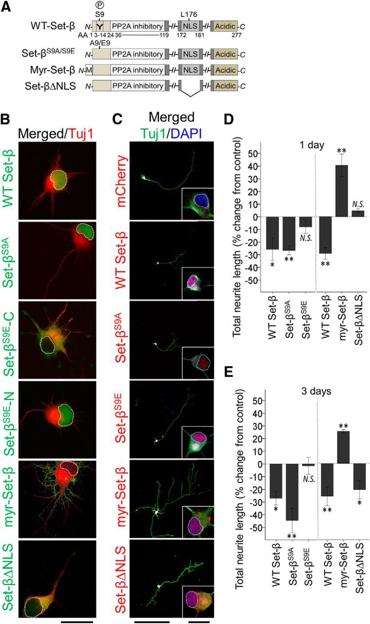
Set-β and Set-β mutants' subcellular localization and effects on neurite growth. A, Set-β domains and mutant constructs, highlighting S9 phosphorylation site (P) and β isoform-specific antibody epitope (Y), PP2A inhibitory domain, NLS, myristoylation (M) tag, and acidic C-terminal domain. B, RGCs transfected with mCherry control or with tagged Set-β constructs as marked, were immunostained at 1 d for Tuj1 (neurite marker, red) and for Set-β tags (green). Set-β, Set-βS9A, and in one third of RGCs Set-βS9E localized to the nucleus (example marked Set-βS9E-N). Set-βΔNLS and one third of Set-βS9E localized to the cytoplasm (labeled Set-βS9E-C). The last third of Set-βΔE localized to both nucleus and the cytoplasm, data not shown. Myr-Set-β localized to cellular membranes and neurites. Scale bar, 20 μm. C, RGCs transfected with mCherry or tagged Set-β constructs as marked, were immunostained at 3 d for fused tags (red), Tuj1 (neurite marker, green), MAP2 (dendrite marker, data not shown), and counterstained with DAPI (nuclear marker, blue). Set-β, Set-βS9A, and Set-βS9E in all cells localized to the nucleus, Set-βΔNLS and myr-Set-β localized to nucleus and cytoplasm, and myr-Set-β also localized to cellular membranes and neurites. Nucleus outlined with dashed white line. Scale bars, 500 μm; insets, 20 μm. D, E, At 1 (D) and 3 (E) days, Tuj1-positive neurites of transfected RGCs were traced. Set-β and Set-βΔS9A significantly suppressed neurite growth, whereas Set-βS9E-N failed to meaningfully suppress neurite growth (N = 3; ≥30 neurons per experiment, mean ± SEM normalized to mCherry shown; *p < 0.05, **p < 0.01 by ANOVA with post hoc LSD). Myr-Set-β significantly increased neurite growth, whereas Set-βΔNLS only affected neurite growth at 3 d (N = 3; ≥30 neurons per experiment, mean ± SEM normalized to mCherry shown; *p < 0.05, **p < 0.01 by ANOVA with post hoc LSD).