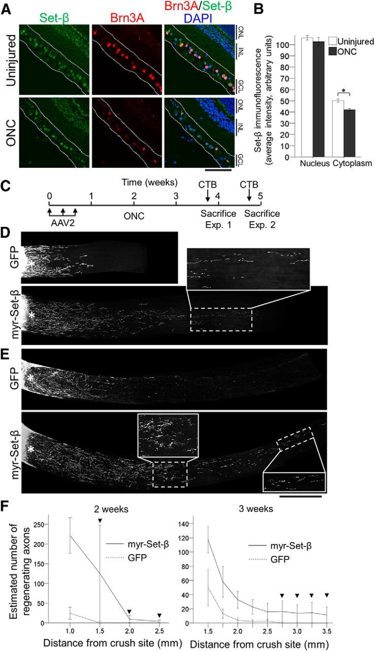
Figure 7.

Myr-Set-β promotes axon regeneration after optic nerve injury in vivo. A, Adult retinal sections from uninjured animals and 5 d after optic nerve crush (ONC) injury were immunostained for Set-β and Brn3A, and counterstained with DAPI (nuclear marker), as marked. Scale bar, 500 μm. GCL, Ganglion cell layer; INL, inner nuclear layer; ONL, outer nuclear layer. B, Analysis of nuclear and cytoplasmic average pixel intensity of Set-β immunofluorescence in injured (ONC) and uninjured RGCs showed that Set-β immunoreactivity mildly decreased in the cytoplasm, although signal intensity was similar in the nucleus. (N = 3; ≥85 randomly selected RGCs per experiment; mean ± SEM shown; *p < 0.05, by ANOVA with post hoc LSD). C, Experimental time-line for optic nerve regeneration experiments with myr-Set-β. D–F, Optic nerves, treated with control GFP or myr-Set-β AAV2 vectors as marked, 2 (D) or 3 (E) weeks after injury. Scale bar, 500 μm; insets, 250 μm (images of merged multiple sections shown). F, Axon regeneration at 2 (left) and 3 (right) weeks after injury demonstrates significantly greater regeneration at increasing distances down the optic nerve. At the longer distances, regenerating axons were only detected in the myr-Set-β-treated retinas (arrowheads; ≥4 animals per group; mean ± SEM shown; p < 0.05 at 2 weeks and p < 0.01 at 3 weeks by ANOVA with repeated measures, post hoc LSD).
