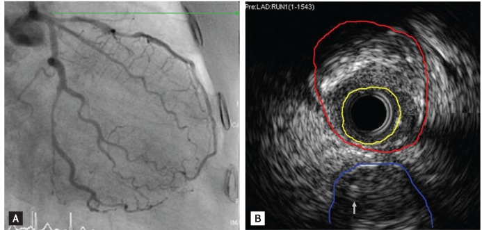
Figure 1.
(A) Initial coronary arteriography shows a 70% to 80% diffuse stenosis in the mid left circumflex artery and a 70% to 80% tandem stenosis in the proximal and mid left anterior descending artery. (B) Intracoronary ultrasound revealed that the minimal luminal area (yellow line) was 2.25 mm2 and the external elastic membrane cross sectional area (red line) was 13.5 mm2 at the ostium. The proximal left anterior descending artery stenotic lesion consisted of mixed plaque. Therefore, the stenotic area was approximately 83.5%. A left circumflex artery (blue line) and a wire (arrow) are shown.

