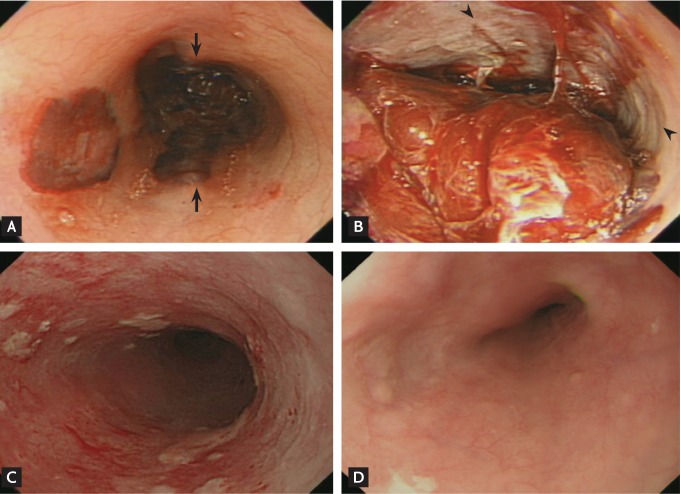
Figure 3.
Serial follow-up endoscopic findings. The initial endoscopy shows (A) black macerated mucosa (arrows) in the mid third of the esophagus and (B) circumferential mucosal necrosis (arrowheads) with a huge adherent blood clot in the distal third of the esophagus. (C) After 1 month, detachment of the necrotic mucosa with re-epithelialization was noted at the same site. (D) Follow-up endoscopy performed 3 months after the first session shows completely healed esophageal mucosa without evidence of ischemic complications.

