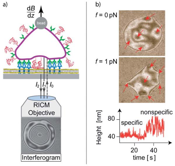
Fig. 2.
Left: schematic view of image formation by reflection interference contrast microscopy (RICM) by interference of light reflected from the cell (I1) and the substrate surface (I2). Lift forces are generated by super-paramagnetic magnetic tweezers subjected to inhomogeneous magnetic fields (dB/dz). Right: interferogram of a test cell adhering to integrin receptors immobilized on the substrate, prior (top) and while applying a lift force of 1 pN (middle). Some initially visible adhesion domains are indicated by arrows. They are revealed by the formation of dark patches in the absence or of sharp edges in the presence of lift forces. The bottom-right panel shows the time evolution of membrane fluctuations during unbinding of an adhesion domain.

