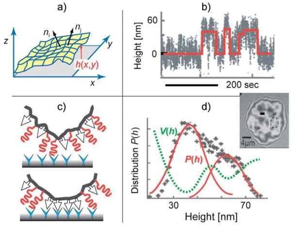
Fig. 4.
Nucleation of CAM–CAM pairs driven by membrane bending excitations. Top-left panel: characterization of the roughness by the local orientation of the membrane normal that is correlated with the coherence length ζp. Bottom-left panel: formation of CAM–CAM clusters by transient displacement of repellers from the site of the CAM–CAM contact. Top-right panel: time sequence of the distance between the membrane from the solid surface h(t) at the particular position (measured by RICM at the site marked by a circle in the interferogram). The red line indicates the random transition of the membrane between the bound state 〈h 〉 ~30 nm and the unbound state at 〈h〉 ~60 nm. Bottom-right: bimodal height distribution Pf(h) defines the double well interfacial interaction potential according to V(h) ∝ kBT ln P(h). Image adapted from ref. 41.

