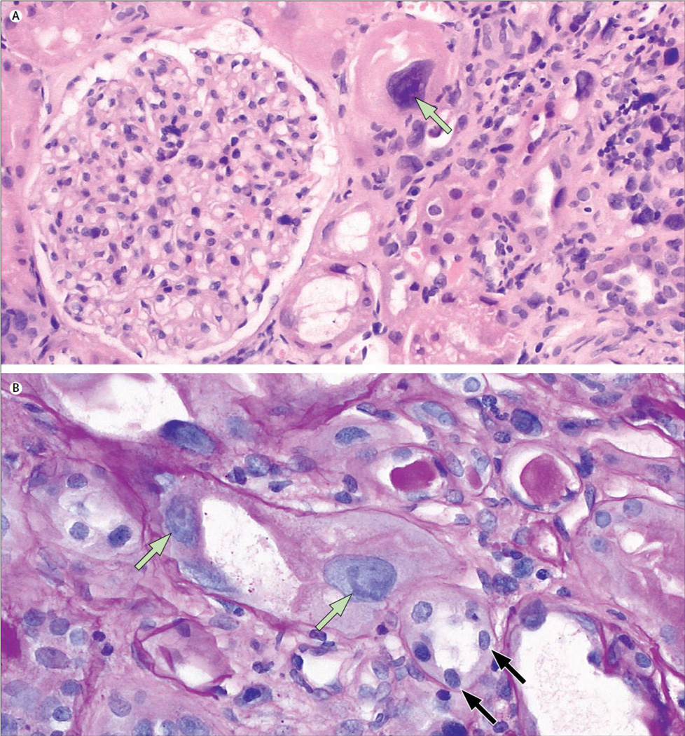
Figure. Light micrographs of renal cells showing karyomegalic interstitial nephritis.
(A) Highly enlarged tubular epithelial cell nucleus (arrow) compared with glomerular nuclei (haemotoxylin and eosin stain, ×200). (B) Karyomegalic cells appear predominantly in proximal tubuli (green arrows). Distal tubular cells are mostly unaff ected (black arrows) (periodic acid-Schiff stain, ×400).

