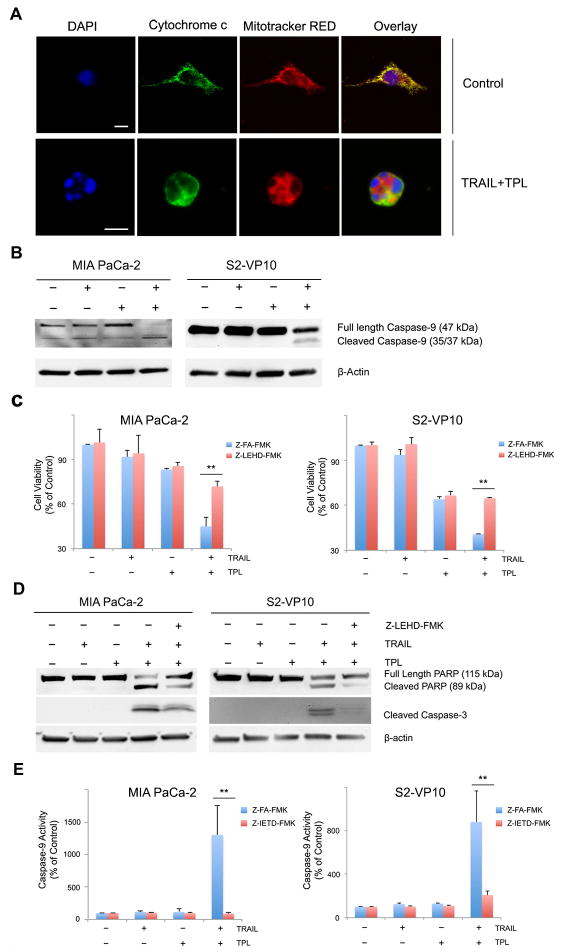
Figure 5. Combination of TRAIL and triptolide induces mitochondrial membrane permeabilization in pancreatic cancer cells.
A. MIA PaCa-2 cells treated with a combination of TRAIL and triptolide for 24h show release of cytochrome c from mitochondrial into the cytosol as evaluated by confocal microscopy. Cytochrome c (green) colocalizes with mitochondrial (mitochondrial red) in a punctuate fashion in control cells, suggesting intra-mitochondrial location. The nuclei have been stained with DAPI (blue). Scale bar, 10 micron/L.
B. MIA PaCa-2 and S2-VP10 cells treated with either TRAIL or triptolide or a combination of both for 24h show cleavage of caspase-9 as evaluated by western blot. β-actin expression was used a loading control.
C. Treatment of MIA PaCa-2 and S2-VP10 cells with caspase-9 inhibitor (Z-LEHD-FMK) leads to rescue of cells from TRAIL/triptolide mediated cell death compared to those treated with a negative control. The bars represent mean ± SEM, n≥3. **p<0.01.
D. PARP and caspase-3 cleavage was assessed in samples treated as described in (C), using western blots. β-actin expression was used as loading control.
E. Treatment of MIA PaCa-2 and S2-VP10 cells with caspase-8 inhibitor (Z- IETD-FMK) leads to decrease in TAIL/triptolide mediated caspase-9 activation compared to those treated with a negative control. The bars represent mean ± SEM, n≥3. **p<0.01.

