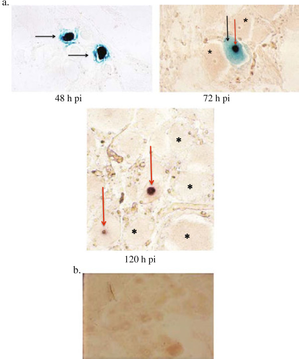
Figure 4.
Detection of AB4Δ75-LacZ and expression of latency associated transcripts (LAT) in trigeminal ganglionic neurons. A. LacZ expression as indicated by X-gal positive cell nuclei representing presence of AB4∆75-LacZ at 48 h and 72 h pi. Two ganglionic neurons with nuclear localization of beta galactosidase activity are shown at 48 h pi (solid arrows). No counter stain, magnification × 400. At 72 h pi, one ganglionic neuron with nuclear localization of beta galactosidase activity (black arrow) and nucleolar localization of LAT (red arrow) is shown. Adjacent neuronal nuclei are negative by both techniques (*). No counter stain, magnification × 400. At 120 h pi, nucleolar expression of LAT only is shown in two ganglionic neurones with nucleolar localization of LAT (red arrow). The nuclei are negative for beta galactosidase activity. Adjacent neuronal nuclei are negative by both techniques (*). No counter stain, magnification × 400. B. No detection of LacZ expression as indicated by X-gal positive cell nuclei or LAT expression in the trigeminal ganglion from an uninfected control pony. No counter stain, magnification × 400.

