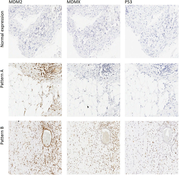
Figure 2.
Immunohistochemistry patterns for MDM2, MDMX and P53. Three distinct patterns of immunohistochemistry staining were identified: normal expression where none of the examined proteins was over-expressed; negative P53 expression with higher scores of MDM2 in comparison to MDMX (Pattern A); positive P53 expression with comparable scores of MDM2 and MDMX (Pattern B).

