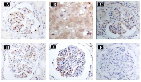
Figure 1.

Immunohistochemical staining. A: HBsAg positive staining in glomerular endothelial cells and mesangial cells in HBV-GN (Magnification of 400x). B: AIM2 positive staining in hepatic cytoplasm in CHB (Magnification of 400x). C: AIM2 positive staining in glomerular endothelial cells and mesangial cells in HBV-GN (Magnification of 400x). D: Caspase-1 positive staining in glomerular endothelial cells and mesangial cells in HBV-GN (Magnification of 400x). E: IL-1β positive staining in glomerular endothelial cells and mesangial cells in HBV-GN (Magnification of 400x). F: AIM2, Caspase-1 and IL-1β negative staining in glomerular endothelial cells and mesangial cells in CGN (Magnification of 400x).
