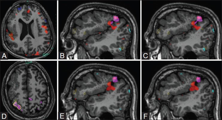
Figure 12(A-F).

Right hemispheric perisylvian polymicrogyria, with preservation of cortical functionality. The left-hand motor task (A) involved repetitive passive clenching of the fingers, and elicited activation in the superolateral aspect of the polymicrogyric cortex (pink blob). Tongue movements (D) generated bilateral cortical activations, with activation located just beneath, and there was a subtle overlap with the left-hand task activation (red blob). (B-C) and (E-F) show overlay images of left hand motor and tongue activations over the polymicrogyric cortex
