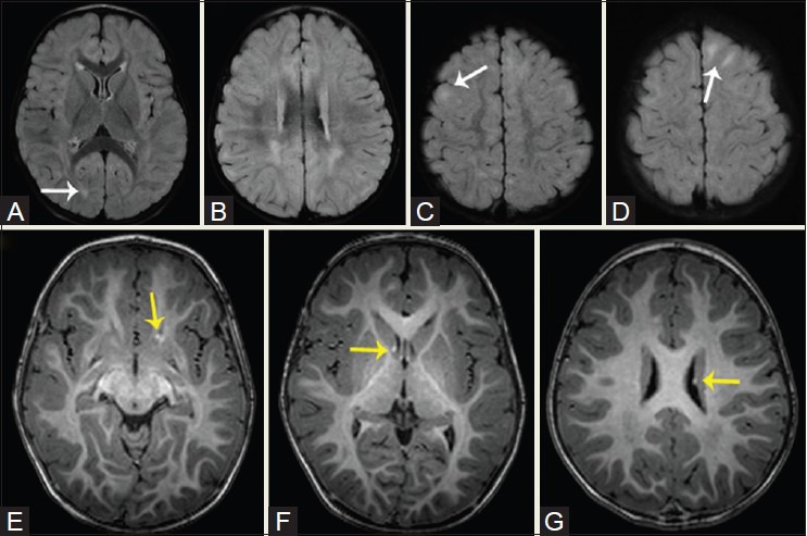
Figure 16(A-G).

A 1-year-old patient with tuberous sclerosis. Axial FLAIR images reveal numerous cortical tubers with subcortical hyperintensity in bilateral frontal and temporal and left occipital lobes. Axial T1W images reveal tiny high-signal subependymal hamartomas around the caudothalamic grooves and margins of the atria of lateral ventricles (yellow arrows)
