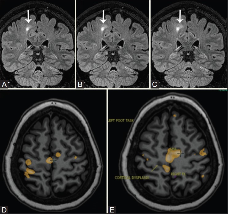
Figure 19(A-E).

A 28-year-old patient with refractory seizures. 3D FLAIR sequence shows focal cortical dysplasia in the medial cortex of the right frontal lobe, just anterior to the pars marginalis. Toe clenching demonstrates activation in right SM1 (yellow blob) immediately postero-medial to the dysplasia, separated by a shallow sulcus. The supplementary motor area [SMA] activation is located anterior to the dysplastic cortex. Finger tapping resulted in activation lateral to the dysplastic cortex in the hand knob (yellow blob)
