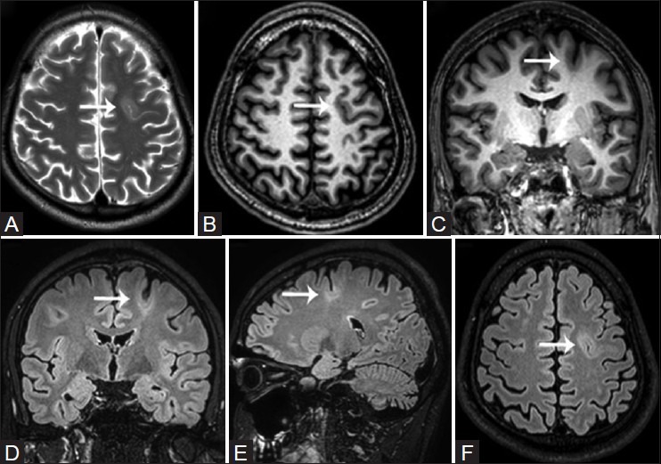
Figure 6(A-F).

Focal cortical dysplasia Type IIB (bottom of sulcus dysplasia) in a 26-year-old patient with complex partial seizures. Cortical thickening with intracortical hyperintense signal and blurring of the cortical-subcortical interphase is visualized along the depth of the left superior frontal sulcus (white arrows). Note a thin comet tail of gray matter coursing through the frontal white matter between the dysplastic cortex and the ventricular margin
