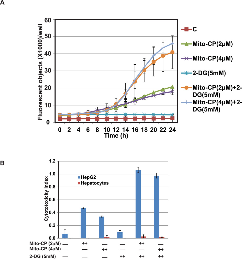
Figure 3.
Mito-CP and 2-DG combination induces cytotoxicity in HepG2 cells determined by YOYO-1 fluorescence assay (A) and cytotoxicity index (B). Measurement of cytotoxicity in real time using YOYO-1 fluorescence in HepG2 cells in response to treatment with 2, or 4 µM Mito-CP and 5 mM 2-DG both individually or in combination for 24 hr (A). YOYO-1 fluorescence increases significantly with combination treatment in HepG2 cells compared to control untreated group as well as treated individually with Mito-CP or 2-DG. Induction of cytotoxicity in HepG2 cells by combination treatment of Mito-CP and 2-DG determined by caspase 3/7 apoptosis assay (B). HepG2 and primary hepatocytes cells were treated with alone or in combination with Mito-CP and 2-DG for 24 hr. and scanned for the fluorescence signal and phase contrast images. Cytotoxicity index in HepG2 cells is highest in cells treated with a combination of 2, or 4 µM Mito-CP with 5 mM 2-DG compared to control treatment. Importantly treatment to primary hepatocytes showed very marginal cytotoxicity index compared to HepG2 cells under the same condition. Average of two independent experiments is shown.

