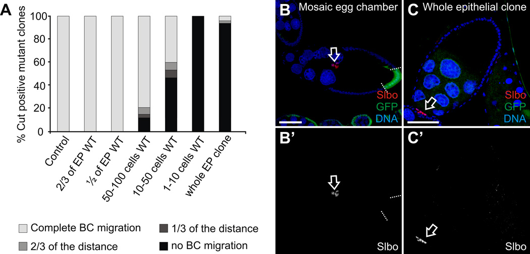
Fig. 5. Quantification of the border cell migration phenotype in the phmFL99 and phmFT59 homozygous mutant egg chambers.
(A) Percentage of the Cut positive mutant clones in the stage 10A egg chambers of the following classes: wild-type (control) egg chambers (n=44); egg chambers with 2/3 of the total FCs being wild-type (n=43); egg chambers with the half of the follicle epithelium being wild-type (n=32); mosaic egg chambers containing 50–100 wild-type FCs (n=33); 10–50 wild-type FCs (n=15); 1–10 wild-type FCs (n=8); and egg chambers containing whole epithelial phm mutant clones (n=46). BC migration in the egg chambers at stage 10A was scored distinguishing the following categories: “completed migration” when BCs reached the oocyte (white); BCs migrated over 2/3 of the distance between the most anterior end of the egg chamber and the border between the nurse cells and the oocyte (light grey); BCs migrated 1/3 of this specified distance (dark grey); and BC cluster failed to migrate and stayed at the most anterior end of the egg chamber, the so-called “no migration” phenotype (black). (B, C) Egg chambers stained with with Hoechst (B, C) to label DNA, and anti-Slbo (B) or anti-Cut (C) antibody to label border cell clusters. Egg chambers are oriented with the anterior to the left. Scale bars represent 40 µm. (B) Stage 10A phm mutant mosaic egg chamber. Mutant cells are marked by the absence of GFP (green), the borders between mutant clones and neighboring wild-type cells are marked by dashed lines. Border cells (open arrow) migrated normally. (C) Stage 10A egg chamber containing phm whole epithelial homozygous mutant clone. Border cells (open arrow) failed to migrate.

