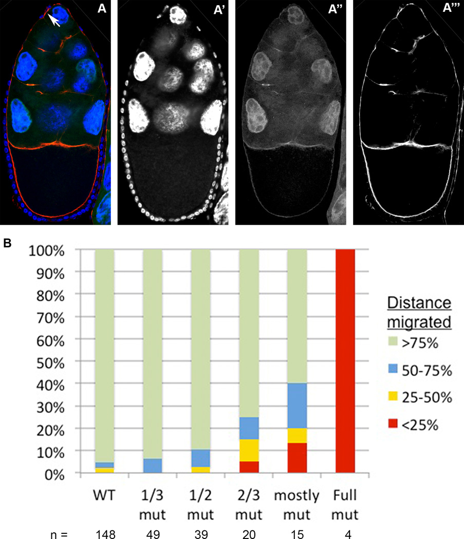
Figure 6. The shd mutant border cell migration phenotype: increasingly larger mutant clones within the follicular epithelium result in more severe migration delays.
(A) A Stage 10 egg chamber with a follicular epithelium that is entirely mutant for shd (observe the lack of GFP in the follicular epithelium in A”), stained with Hoechst to label nuclei (A’), and phalloidin to label cell membranes (A”’). Anterior is oriented upward. The border cell cluster, indicated with a white arrow in A, fails to detach from the epithelium at the anterior of the egg chamber. (B) Quantification of border cell migration behaviors in egg chambers with epithelial clones of increasingly larger proportions. The size of each colored bar represents the percentage of egg chambers in which border cells have migrated through each quadrant of the wild type migration distance. Egg chambers with increasingly larger sized epithelial clones exhibit more severe delays in migration.

