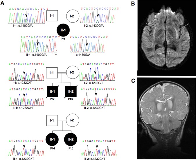
Figure 1.
Pedigrees and radiological features. A: Pedigrees and electropherograms of the MTO1 genomic region encompassing the nucleotide substitutions in patients and available parents. Black symbols designate affected subjects. B: Brain MRI of Pt1. Transverse FLAIR image showing abnormal hyperintensity in the region of the claustrum and surrounding capsulae (arrows). C: Brain MRI of Pt2. Coronal T2-weighted sequence showing abnormal hyperintense signals of the thalami and diffusely abnormal signal in the subcortical white matter. Lesions are also present in the brainstem. The cerebellar folia are normal.

