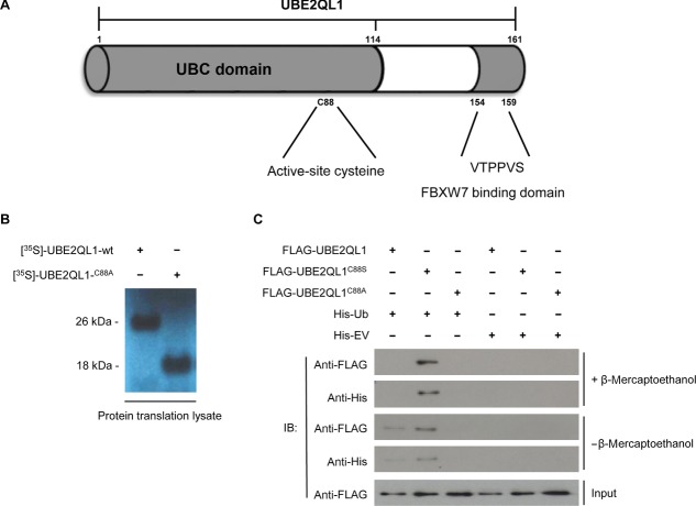
Figure 4.
UBE2QL1 binds monoubiquitin in vivo. A: Schematic diagram of UBE2QL1 showing the ubiquitin conjugating (Ubc) domain containing an active-site cysteine at residue 88 and a FBXW7 recognition motif (residues 154–159). B: Transcription/translation lysate system produces wtUBE2QL1 at ∼26 kDa and UBE2QL1C88A at ∼18 kDa suggests wtUBE2QL1 is monoubiquitinated (ubiquitin Mr 8.5 kDa). C: HEK-293 cells were transfected with either FLAG-tagged UBE2QL1-wt or UBE2QL1-C88S or FLAG-UBE2QL1-C88A mutants and either His6-tagged ubiquitin (His-Ub) or empty vector (His-EV). His pulldown followed by immunoblot (IB) analysis demonstrated that UBE2QL1-wt is monoubiquitinated in vivo (bands at Mr of ∼26 kDa). Input levels of UBE2QL1-wt, UBE2QL1-C88S, and UBE2QL1-C88A in the cell lysate are indicated (bands at Mr of ∼18 kDa).

