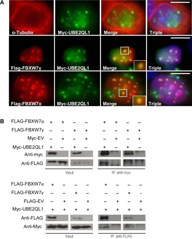
Figure 5.

UBE2QL1 colocalizes and immunoprecipitates with FBXW7. A: HeLa cells were transfected with either myc-UBE2QL1 alone and stained with antibodies against α-tubulin (red) and myc (green), which showed a nuclear localization of UBE2QL1 (upper panel). When cotransfected with myc-UBE2QL1 and either FBXW7 γ (middle panel) or FBXW7α (lower panel) and stained with antibodies against FBXW7α or FBXW7γ (red) and myc (green) there was nuclear colocalization of UBE2QL1 with FBXW7α and FBXW7γ. Triple refers to DAPI nuclear staining (blue) and the merged images together. B: HEK-293 cells were transfected with either myc tagged to an empty vector (myc-EV) or myc-UBE2QL1 and FLAG-FBXW7 as indicated. Immunoprecipitation (IP) of myc-UBE2QL1 (upper panel) followed by immunoblot (IB) analysis with antibody against the FLAG tag identified FBXW7α and FBXW7γ as UBE2QL1 interacting proteins. The reciprocal experiment whereby IP of FLAG-FBXW7α and FLAG-FBXW7γ (lower panel) followed by IB analysis with antibody against the myc tag also identified FBXW7α and FBXW7γ as UBE2QL1 interacting proteins. Input levels of FBXW7α, FBXW7γ, and UBE2QL1 in the cell lysate are indicated.
