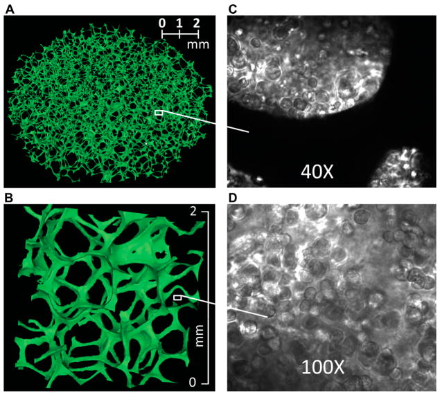
Figure 1.
Images of a Cytomatrix™ carbon scaffold. (A) Three-dimensional rendering of a μCT image of the Cytomatrix™ carbon scaffold. (B) Magnification of a 2 × 2 × 1 mm3 portion of the scaffold. (C) Light microscope image of human MDA breast cancer cells suspended in Matrigel™ and then cultured in a Cytomatrix™. (D) Magnified view of human MDA breast cancer cells within a Cytomatrix™.

