
Figure 3.
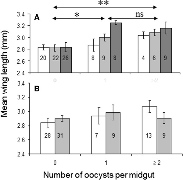
Size of Anopheles gambiae (A) and Anopheles stephensi (B) mosquitoes infected at different intensities by oocysts of Plasmodium yoelii nigeriensis. Differently coloured bars represent replicates ± SEM. Sample sizes are shown within the bars.