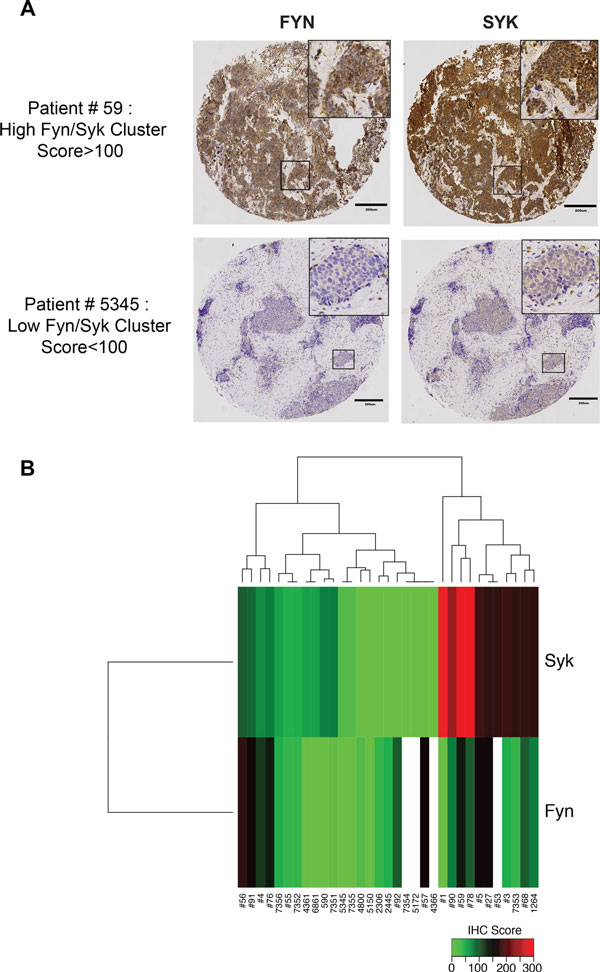
Figure 4.
Co-expression of SYK and FYN in a subset of SCLC tumors. Contiguous sections of TMAs from 39 SCLC patient specimens were stained with antibodies to SYK and FYN, respectively. Stained sections were scored by a pathologist as described in Methods. (A) Representative stained sections showing positive (upper) or negative (lower) results. See text for additional details. Tumor spot images were captured by brightfield microscopy at 20X magnification. (B) Unsupervised hierarchical clustering heatmap of SYK and FYN immunostaining intensity scores across SCLC patients distinguished positive from negative tumors as described in Methods. Red and green indicate high and low expression, respectively. Specimens that are positive for both SYK and FYN segregate in one cluster, on the right. Patient ID shown below the heatmap.

