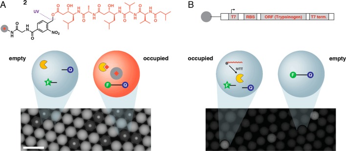
Figure 4.

Bead-based assays in picoliter-scale droplets. (A) UV-photocleavable pepstatin A (1, red) attached to TentaGel resin beads (gray circle with red diamond) were distributed into droplets along with HIV-1 protease (yellow), fluorogenic HIV-1 protease substrate (F-Q), and internal standard. After UV exposure, bead-occupied droplets contain significant pepstatin A concentration (red droplet), inhibiting proteolytic F-Q digestion. In empty negative control droplets, F-Q proteolysis is uninhibited (blue droplet). (B) Magnetic beads functionalized with trypsinogen gene were distributed into droplets containing IVTT reagent, enterokinase, and a fluorogenic trypsin substrate (F-Q). In occupied droplets, trypsin expression leads to substrate proteolysis and fluorescence. No trypsin is expressed in empty droplets, and the substrate remains quenched. Scale = 100 μm.
