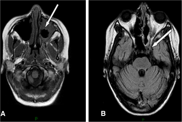
Figure 1.

Magnetic resonance imaging of head. A. Postgadolinium T1-weighted axial magnetic resonance image showing nonenhancing tissue (arrow) in the left nasal cavity and periorbital site compatible with necrotic mucosa. B. Similar technique showing note semiocclusion of the left internal carotid artery (arrow) and thrombosis of the right cavernous sinus, although there was no radiographic evidence of cerebral infarction.
